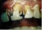
 Clinical note: The oral
cavity has a large and varied bacterial flora. Bacteria may
accumulate and form a layer (plaque) on a tooth, releasing acids
that demineralize the enamel and dentine and produce dental
cavities. Other bacteria proliferating in the gingival cleft may
destroy the periodontal ligament, cause resorption of alveolar bone,
and loosening of teeth. Clinical note: The oral
cavity has a large and varied bacterial flora. Bacteria may
accumulate and form a layer (plaque) on a tooth, releasing acids
that demineralize the enamel and dentine and produce dental
cavities. Other bacteria proliferating in the gingival cleft may
destroy the periodontal ligament, cause resorption of alveolar bone,
and loosening of teeth.Examine
a section of developing tooth in the lower jaw of the fetal skull on
slide 130. The stage shown is closest to that in Fig. 15-11.
Identify the dental pulp, odontoblasts, dentine, enamel (partially
gone), and the (partially disrupted) layer of ameloblasts (Fig.
15-8 through 15-11).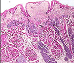
Briefly describe how the
odontoblast and ameloblast layers form the tooth.
Examine the preserved-mounted
specimen of tongue and larynx,
- Note the macroscopic features
(median sulcus, circumvallate papillae, filiform papillae,
lingual tonsils, etc.)
What is the functional
significance of these various surface structures on the tongue?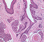
Examine sections of the tongue
(slides 70 and
77) and identify filiform
and fungiform papillae on
the dorsal lingual surface (Figs. 15-4 and 15-5). On
slide 77 look
for a circumvallate papilla. (Not every
slide 77 shows such a
papilla, so you may have to share your neighbor's slide).
- Identify taste buds and the
serous von Ebner's glands associated with circumvallate
papillae (Figs. 15-5 and 15-6).
What are the chief histological
differences among the different types of lingual papillae?
Let's take a look at the
salivary glands. |