Stomach --
 site
where food is converted to a thick fluid for most efficient
enzymatic digestion of macromolecules. site
where food is converted to a thick fluid for most efficient
enzymatic digestion of macromolecules.Examine a section
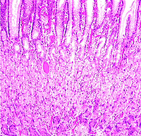
of the fundic region of the stomach (slide 101).
- Identify first the layers and
sublayers (Fig. 15-15), noting that the simple columnar
epithelium invaginates into gastric pits, which are tightly
packed together side-by-side
- These lead into the gastric
glands which extend down all the way to the thin muscularis
mucosa. Cells of the lamina propria are seen scattered loosely around the
gastric pits.
Why is the lamina propria of
the stomach more difficult than usual to study histologically?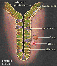
On the same slide (101), find a
region of mucosa in which the gastric glands are cut longitudinally.
Starting at the surface, identify the mucus-secreting cells (called
surface and neck cells depending on their proximity to the surface
lining the stomach cavity), the large eosinophilic parietal cells,
and the smaller, more basophilic peptic cells (Figs. 15-18 through
15-24). (Image
courtesy of GERD Information Resource
Center)
How would you classify the
gastric glands morphologically?
Why are the products of all
three of these cells critical for proper digestion of food in the
stomach?
Using electron micrographs in
the atlas, study the ultrastructural features of peptic cells (Fig.
15-24) and parietal cells (Fig. 15-22).
What highly unusual
structures of parietal cells make them uniquely suited for their
function?
On to the
pyloric glands and small intestine. |