 Examine
the mucosa of the pyloric stomach (Fig. 15-19), Examine
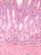
the mucosa of the pyloric stomach (Fig. 15-19),
- Note that the pyloric glands,
unlike the gastric glands found in the fundus and body of the
stomach, contain mainly mucus-secreting cells and few parietal
cells.
- Plus a few scattered gastrin
cells representing the diffuse endocrine system along the gut.
- Cells of the enteroendocrine
system will be considered more fully in a later topic, but they
are not well-shown in routinely stained slides.
Compare and contrast the
pyloric glands with the gastric glands.
Small Intestine -- site where
digestion is completed, using enzymes from the pancreas and bile,
and where products of digestion are absorbed. The three regions
(duodenum, jejunum, and ileum, are similar histologically.) Examine
the plastinated segment of small intestine, noting the features of
its wall and mucosal folds.
Two sections of the duodenum (slides
4,
50), one section of jejunum (slide 158), and two of ileum (slides
21,
37) should be studied.
- On slides
4,
21, and
158,
identify the major layers of the small intestine and the tightly
packed together villi formed as projections of the mucosa
layer (Figs. 15-25 through 15-35).
- Plicae circulares, large folds
of the mucosa and submucosa (Fig. 15-26), are visible on slides
37 and
50, but are best shown on slide
158.
What is the difference between a
villus and a plica circulares?
Brunner who? |