 Parathyroid
gland (Fig. 20-22) -- secretes the most important hormone regulating blood
calcium levels. Parathyroid
gland (Fig. 20-22) -- secretes the most important hormone regulating blood
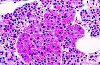
calcium levels.Examine a
section of parathyroid gland (slide 69). Note the very thin capsule
and fine septa (Fig. 20-23) and identify the clumps of
chief (principal) cells which secrete PTH and of oxyphil cells which
are packed with large mitochondria but whose function remains
unknown. (Some examples of slide 69 also have thyroid and thymus
tissue.)
Explain how the parathyroid glands
are related functionally and embryologically to the parafollicular
cells of the thyroid gland.
Adrenal gland -- consists of
two endocrine tissues (cortex and medulla) that are functionally distinct
(Fig. 20-14).
Examine adrenal glands on slides
20,
75 and
143. These are flattened glands with concentric layers of secretory tissue (Fig.
20-14). In the distinct capsule associated
with adipose tissue, notice the small arteries entering the gland
and the vascular plexus just inside the capsule (Fig. 20-15).
On the same two slides (20 and
143),
examine the cortex and identify the three layers or zones in which
the steroid-secreting cells have slightly different arrangements,
with groups of cells separated by fine, well-vascularized support
tissue septa. The outermost zona glomerulosa (Fig. 20-15) has cells
secreting mineralocorticoids arranged in irregular rounded clumps ("glomeruli").
The middle and widest layer, the zona fasciculata (Fig. 20-15), has
cells making glucocorticoids arranged in strands ("fascicles"). The
innermost layer, the zona reticularis (Fig. 20-15), shows irregular,
branching ("reticular") cords of cells secreting small quantities of
sex hormones (androgens).
Study the electron micrographs of
steroid-secreting cells in Fig. 20-13.
Why do steroid-secreting cells
stain poorly in routine light-microscope staining procedures?
Addison's
disease and beyond. |