 Testis
and spermatogenesis - Examine the preserved-mounted specimens of
testes and vas deferens, noting the macroscopic features (lobes,
tubules, etc.) for correlation with these structures on the slides. Testis
and spermatogenesis - Examine the preserved-mounted specimens of
testes and vas deferens, noting the macroscopic features (lobes,
tubules, etc.) for correlation with these structures on the slides.
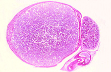
Examine both the trichrome-stained
section of testis (slide 55) and the H&E stained testis (slide
119). Identify the capsule (tunica albuginea), interstitial tissue
or stroma (which is not very well preserved in
slide 55), and the seminiferous tubules (Fig. 18.3 and 18.4).
What constitutes the parenchyma
of the testis?
Clinical note: Failure of the
testes to descend into the scrotum during fetal development
(cryptorchidism) maintains their temperature at 37oC which inhibits
spermatogenesis, although testosterone production can still occur.
Excessive blood flow or dilated vasculature in the scrotum (varicocoele)
is another potential cause of male infertility and can be surgically
corrected.
Study the schematic diagram (next
page) of spermatogenesis and its relationship to the sustentacular
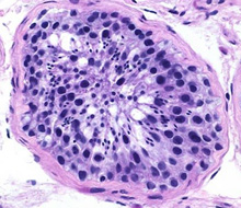
Sertoli cells. On slide 155 examine the cells inside a seminiferous
tubule and distinguish their interrelationships during
spermatogenesis. In a tubule cut transversely, identify myofibroblasts (myoid cells), Sertoli cells (with nucleoli),
spermatogonia, and the large primary spermatocytes (Fig. 18.5). The
smaller secondary spermatocytes are much more short-lived and
therefore more difficult to find. (Do not spend time looking for
these.) Identify spermatids and differentiating spermatozoa. Try to
distinguish some of the morphological stages and changes that occur
in the differentiating spermatids during spermiogenesis (Fig. 18.6)
and study the ultrastructure of a mature spermatozoon (Fig. 18.7).
Describe the process of meiosis
and the cellular changes during spermatogenesis.
Sketch the changes a spermatid
undergoes during spermiogenesis.
More about the
testis. |