 Most
cases of testicular cancer (95%) are of germ cell origin. The rest
are derived from Sertoli cells or Leydig cells. Examine the electron
micrographs of Sertoli cells (Fig. 18.8). Most
cases of testicular cancer (95%) are of germ cell origin. The rest
are derived from Sertoli cells or Leydig cells. Examine the electron
micrographs of Sertoli cells (Fig. 18.8).
Explain the relationship of Sertoli
cells to developing germ cells and the nature of the "blood-testis
barrier."
On slides
119 and
155, examine the
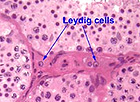
interstitial tissue between tubules and identify the large
interstitial cells (Leydig cells) (Fig. 21-4).
 What
is the function of Leydig cells and what hormone stimulates this
function? What
is the function of Leydig cells and what hormone stimulates this
function?
Back on
slide 55, look at other
regions of the section and identify the rete testis, in dense
connective tissue but having an irregular arrangement of spaces
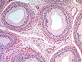
lined by simple cuboidal epithelium (Fig. 21-9). On
slide 40
identify the efferent ductules, loose tubules again lined with tall
ciliated epithelial cells (Fig. 21-10).
What happens in the rete testis
and how does the epithelial lining accomplish this?
What is the function of the
cells lining the efferent ductules?
The epididymis is a long
convoluted duct with smooth muscle. Examine the beginning or head of
the epididymis and adjacent efferent ductules on
slide 40 and the
tail of the epididymis (leading to the vas deferens) on
slide 39.
Identify the tall columnar or pseudostratified epithelium with
stereocilia and the stored spermatozoa in the lumen (Fig.
21-11).
What apical structures are found on
the columnar cells lining the epididymis and what is its function?
Now for the vas
deferens and seminal vessicles. |