 Mammary
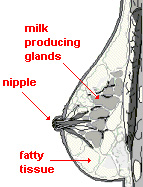
Gland (Fig. 22-23) -- considered functionally to be part of the female
reproductive system. Mammary
Gland (Fig. 22-23) -- considered functionally to be part of the female
reproductive system.Examine
the diagram of the mammary gland, noting the lactiferous ducts and
sinuses which converge at the nipple (Fig. 22-23).
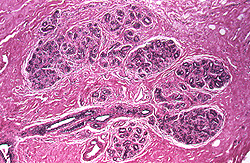
Examine a section of an active
mammary gland from a pregnant,
woman (slide 108).
- At low power, identify the fibrous
support tissue, larger branches of lactiferous ducts,
plasma cells, and adipose
tissue and lobules of secretory acini (Fig. 22-24 and 22-25).
- At higher power, examine the
 epithelium of the secretory acini (or alveoli) of cells with round
nuclei and the surrounding myoepithelial cells with more
oval-shaped nuclei (lacking nucleoli), but inside the basement
membrane (Figs. 22-25 and 22-26).
What type of epithelium lines
the lactiferous ducts?
How does the breast tissue on
this slide differ from mammary tissue before puberty?
More about the
breast. |