 Specialized
connective tissue Specialized
connective tissueExamine
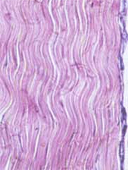
the appearance of tendon (Fig. 5-22) ("dense regular" CT) in
the demonstration slide.
- Note the regular parallel
arrangement of the collagen fibers and relative absence of other
cells besides fibroblasts.
- The relatively low density of
cells and blood vessels among the collagen bundles in tendon
helps to increase the tensile strength of this tissue, but also
contributes to its extremely slow rate of healing after injury.
 What
is the functional advantage for this type of fiber arrangement? What
is the functional advantage for this type of fiber arrangement?
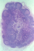
Examine the reticulin fibers (Fig.
5-12) in the silver-stained, oval lymph node on slide
99.
- Also note the appearance of the
collagen bundles in the CT surrounding the lymph node, comparing
it with that of the stains examined earlier.
What is the composition of
reticulin fibers and their function in lymphoid tissue?
Now let's look for
elastic fibers and adipose tissue. |