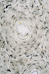
Examine the two samples of compact,
lamellar bone on slide 32.
- This unstained tissue was not
decalcified and was prepared as thin wafers using a grinding
stone rather than a microtome.
- Cells are not preserved by this
method, but the lamellae, lacunae, and canaliculi of osteons (Haversian
systems) are shown very well (Figs. 8-9 and 8-10).
- Study both specimens on
slide 32
for longitudinal and transverse orientations of osteons.
Identify Haversian canals and if possible Volkmann’s canals,
connecting channels between neighboring osteons.
Draw a group of 3 osteons cut
transversely, showing all the structures listed above.
What structures occupied the
various cavities of osteons in the living tissue?
What is the significance of the
irregular "interstitial systems" seen in transverse specimen?
Clinical note: The normal
balance between bone deposition and absorption is influenced by
steroid hormones and in older individuals this balance is disturbed
and there is slow, progressive reduction in bone mass per unit
volume. This occurs in both sexes but is accelerated in women after
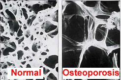
menopause and often leads to osteoporosis (image at right),
in which the reduction in mass of cortical bone and the number of
trabeculae makes the bones fragile.
Now let's consider
bone and joint formation. |