Synovial joint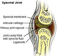
Examine the microscopic section of a
synovial joint that is on demonstration, carefully identifying all
the elements labeled in Figs. 8-19 through 8-21.
- Note that in this slide the
joint may be too immature to allow specific articular cartilage
to be clearly distinguished as shown in the figures.
- Pay particular attention to the
synovium, or synovial membrane, which is diagnostic for this type of joint.
What cartilage type is the
articular cartilage and what is its function?
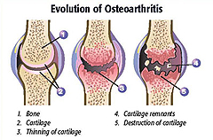 Clinical note: Articular
cartilage normally begins to break down slowly as we age,
potentially leading to inflammation associated with osteoarthritis.
This process can be inhibited by regular exercise and full use of
the joints because recurrent pressure on the cartilage and other
parts of joints improves tissue maintenance. Injection of hyaluronan
solution into the synovial cavity is a routine treatment for severe
arthritis. Dietary supplementation with chondroitin sulfate and
glucosamine has also been shown to slow progression of arthritis.
Click the image for a bigger view. Clinical note: Articular
cartilage normally begins to break down slowly as we age,
potentially leading to inflammation associated with osteoarthritis.
This process can be inhibited by regular exercise and full use of
the joints because recurrent pressure on the cartilage and other
parts of joints improves tissue maintenance. Injection of hyaluronan
solution into the synovial cavity is a routine treatment for severe
arthritis. Dietary supplementation with chondroitin sulfate and
glucosamine has also been shown to slow progression of arthritis.
Click the image for a bigger view.
Why might you expect dietary
supplementation with glucosamine and chondroitin sulfate to possibly
help maintain cartilage and delay arthritis?
Intervertebral joints
- Examine Fig. 8-22
and the fibrocartilage of slide 131. Understand the relationship
to the fibrocartilage and the periosteum of the vertebral bones.
What exactly is a herniated or
“slipped” disk?
Why is such a condition often
painful?
Now let's consider
blood cells and hemopoiesis. |