Intramembranous Ossification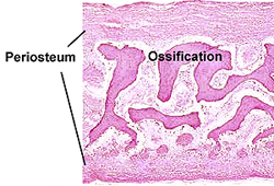
- Use Fig. 8-13 to locate regions
of intramembranous bone development in the sections of fetal
rodent head on slide 130.
- Identify the periosteum and as
many of the bone cells as possible.
What is the difference between
osteoid and woven bone?
Endochondral Ossification
- Locate the cartilaginous region
on slide 34. (This is not a
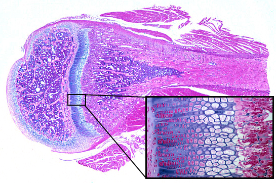 typical epiphysis of a joint, like
that shown in Junqueira's presentation of endochondral
ossification.) typical epiphysis of a joint, like
that shown in Junqueira's presentation of endochondral
ossification.)
- On the slide carefully study all
areas of the growth plate, as shown in Figs. 8-14 through
8-17,
moving from the cartilage to the diaphysis of the bone.
Click to enlarge image.
- Concentrate on the chondrocytes
and matrix, noting the changes in these structures as you move
through the various functional zones.
- zone of reserve cartilage
- zone of
chondroblast proliferation
- zone of chondrocyte hypertrophy
- zone of cartilage
calcification and chondrocyte degeneration
- zone of osteogenesis
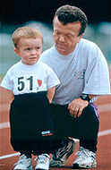 |
Clinical note: Achondroplasia
is an inherited disorder in which there is reduced proliferation of
the chondrocytes in the epiphyseal plate of long bones. The result
is a form of dwarfism in which the trunk is of normal length, but
the extremities are short. Describe in your own words the
steps by which cartilage is formed and then replaced by bone during
this developmental process. (This is important.)
Now for
joints. |
|