Unlike the ground bone specimens
studied in the last lab, the specimens of two small bones on
slide
34 and the bone on slide 129
and 104 were decalcified chemically and then
mounted and sectioned with a microtome.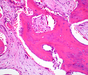
- After staining with H&E all the
cells are readily distinguishable.
-
Bone
Slide 129
and 104shows osteons very
well.
-
Slide 34 is mostly
cancellous bone, but a small area of compact bone is like that
of Fig. 8-7.
- Find this area and compare
the appearance of the osteons to those you saw in the ground
specimen on slide 32.
- The rest of the bone tissue
present on slide 34 is cancellous bone (Fig.
8-7) and one
specimen was undergoing fracture repair. There is much to
identify and study on this slide.
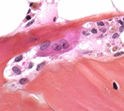 Locate and carefully study the
following structures on slide 34: Locate and carefully study the
following structures on slide 34:
- Active and inactive periosteum
(Fig. 8-6),
- Marrow (Fig. 8-6 and 8-8),
- Osteoblasts and osteoid
(Fig. 8-2 and 8-3),
- Multinucleated osteoclasts which
may be in Howship’s lacunae (Fig. 8-5), and
- The very delicate layer of
endosteum that separates the boney trabecula from the marrow
(Fig. 8-6).
What are the functional
relationships among the periosteum, osteoblasts, osteoid, osteocytes,
and osteoclasts?
How are these relationships
important in the process of bone repair?
Ossification
centers. |