Skeletal or Striated Muscle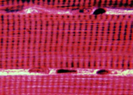
The most characteristic features of
skeletal muscle are large, multinucleate fibers with
cross-striations.
Examine the musculature of the larynx
(slide 7) using low power and study its organization into fascicles.
- Identify epimysium and
perimysium.
- With the 40X objective, examine
muscle fibers cut in transverse and in longitudinal section and
identify endomysium and blood vessels of various sizes (Figs.
10-3 and 10-4).
Where are the nuclei of these
muscle fibers located? Nuclei of what three cell types are located
just outside the muscle fibers?
Clinical note: Exercise or increased use of specific
muscles produce hypertrophy or increased fiber size, while disuse
results in muscle atrophy. One form of the hereditary disease
muscular dystrophy, in which fibers do not mature properly or
survive, results from a defect in the gene for dystrophin, a large
protein with poorly understood functions located just inside the
sarcolemma.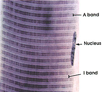
Examine the striations on
longitudinally cut fibers, comparing them with Figs. 10-7 and 10-8.
- Identify A bands and I bands and
- Be sure you understand what
these bands represent in terms of sarcoplasmic organization
(Figs. 10-8 through 10-12).
Based on the diagrams of Fig.
10-12, would you say the muscle on slide 7 was relaxed or contracted
when it was fixed? Why?
Examine the transversely cut fibers
on slide 7 again. Try to distinguish individual myofibrils. Compare
what you see to the EMs and the diagram in Fig. 10-8.
Draw what the myofilament
arrangement in a sarcomere cross-section would most likely be in a
relaxed muscle.
Electron
microscopic images |