 Study
the electron micrographs (Figs. 10-7c and 10-10) and diagrams (Fig.
10-8 and 10-11). Study
the electron micrographs (Figs. 10-7c and 10-10) and diagrams (Fig.
10-8 and 10-11).
- Note particularly the
organization around myofibrils of mitochondria and the various
components of the conducting system for contractile stimuli,
i.e., the T tubules and the sarcoplasmic reticulum.
What exactly are "myofibrils"
and why is the precise organization of the "conducting system"
around them so important?
Examine the musculature of the tongue
on slides 8 and
77.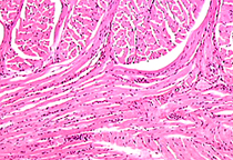
- Identify on each the various
features just studied on slide 7, compare their appearance when
stained by Masson trichrome (slide 8) and H&E (slide 77).
Sketch and label three
“regions” of connective tissue that show up particularly well in
muscle stained with Masson’s trichrome.
Examine developing skeletal muscle of
the fetal tongue and face (slide 159).
- Identify the fusing myoblasts
and the fused but not-yet-striated myotubes as shown in
the diagram of striated myogenesis (Fig. 10-2).
Compare and contrast myoblasts
and satellite cells with regard to histology and function.
Moving right along to
smooth muscle. |