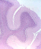
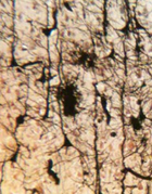
 Before beginning the study of the
slides for this lab, examine the preserved-mounted specimens of CNS
and PNS tissue, noting especially the gross or macroscopic
appearance of the tissues (cerebrum, cerebellum, meninges,
perineurium, etc.). The bigger view will give you a heads up on what
you will be looking for on the slides. Before beginning the study of the
slides for this lab, examine the preserved-mounted specimens of CNS
and PNS tissue, noting especially the gross or macroscopic
appearance of the tissues (cerebrum, cerebellum, meninges,
perineurium, etc.). The bigger view will give you a heads up on what
you will be looking for on the slides.
Cells and general organization of the
Central Nervous System (CNS)
- Looking at
slide 112 and
slide 125, and
preserved-mounted specimens with the eye alone, note the
difference between white and gray matter.
- Then examine a section of the
cerebral cortex (slide 112) and identify
- neuropil,

- neurons,
- oligodendrocytes, and
- astrocytes (Fig. 9-9).
- Study the diagram of neurons (Fig.
9-3).
- On
slide 112, identify
nucleoli and chromatophilic Nissl substance in neurons and understand their
significance.
Why is a third type of glial
cell, the microglial cell, relatively rare?
On the cerebral cortex section of
slide 112, examine the edge showing remnants of the innermost
meningial layer (pia mater) and note the intracerebral
penetration of blood vessels from this surface. Compare this with a
similar area of vessel penetration into the spinal cord as shown in
Fig. 9-18.
Pia mater covers arteries and veins
in the brain. Other components of the meninges are the arachnoid
and the dura (Fig. 9-19). Note their interrelationship in the
diagram.
Clinical note: The
meninges are the site of various medical problems, such as
meningitis, subdural hematomas, subarachnoid hemorrhage, and
meningiomas.
Cerebellum
and spinal cord. |