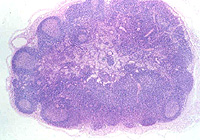
 Lymph
nodes Lymph
nodesExamine sections of
lymph nodes on slides 30, 79 and
42 and note first the overall
organization and the specific structures shown in Figs. 14-17
through 14-19.
Identify
- The cortex,
- Paracortex,
- Medulla
- Subcapsular sinus (Fig.
14-19)
- The large clusters of cells, the
lymphoid follicles with germinal centers (Fig. 14-18).
- Various cells with different
immune functions are located in these follicles.
Examine the specimen stained
immunohistochemically in Fig. 14-20 and note the different
localizations of T cells and B cells.
 Did
the cortex of the thymus show follicles of lymphocytes? Did
the cortex of the thymus show follicles of lymphocytes?
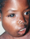
Clinical note: Lymphomas
involve neoplastic growth of lymphocytes, commonly within lymph
nodes. There are several types, depending on the type of lymphocyte
and the stage of differentiation involved. In the early stage,
lymphomas commonly involve cell accumulation caused by their failure
to undergo apoptosis. Pictured here is a child with Burkitt's
lymphoma.
The silver-stained specimen on
slide
99 shows very well the reticular network and other supporting
tissues, as well as the sinuses of the lymph node (Fig. 5-12b).
Identify lymph.
- Can afferent or efferent
lymphatics be identified in this section?
What are some of the unique
staining characteristics of the stain used on
slide 99?
A little more on the
structure of lymph nodes. |