 Cells
in Lymph and the thymus Cells
in Lymph and the thymus
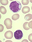
Examine the blood smear slide (10) again, focusing attention only on
the structure of the lymphocytes of different sizes.
What technique could you use to distinguish T and B
lymphocytes on a slide?
What are the respective
functions of T lymphocytes and B lymphocytes?
Thymus
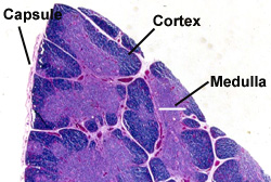
 Examine
a section of thymus (slide 126), noting Examine
a section of thymus (slide 126), noting
- The capsule,
- Septa, and
- The organization of lobes into a
basophilic cortex and eosinophilic medulla (Fig.
14-10).
Identify the epithelial-reticular
cells or epitheliocytes (Fig. 14-11) which make up the framework of
the thymus.
- These are larger and paler than
the lymphocytes. In the
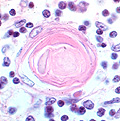
 medulla,
identify the masses of epithelial cells, thymic or Hassal's
corpuscles (Fig. 14-10 and 14-12 ). medulla,
identify the masses of epithelial cells, thymic or Hassal's
corpuscles (Fig. 14-10 and 14-12 ).
Examine also a section of involuted
thymus (slide 123) and note
the fatty infiltration and lymphocyte depletion.
What are three functions of
thymic epithelial cells?
Identify macrophages in
the thymic cortex (they are typically larger and more pale than
lymphocytes and less numerous than the epithelial-reticular cells) and look for mitotic figures among the lymphocytes of
this region.
What are some functional
differences between lymphocytes in the thymic medulla and cortex?
Next let's look at a
lymph node. |