During this lab examine the
preserved-mounted, hemisected heart with parts labeled, and the
preserved specimens of arteries and veins, noting the macroscopic
features of each.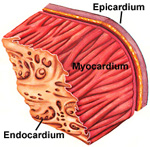
Heart -- Two microscopic
specimens of the walls of heart ventricles are on slides
64 and
65,
stained with periodic acid-Schiff reagent (PAS) and with Masson trichrome respectively.
Slide 65 has some staining artifacts
and folds, but shows the blood vessels and dense CT in the
myocardium well. Examine the major features of the heart on both
slides. An H&E stained heart specimen is on
slide 139.
- Identify the three layers of the
heart wall (Fig. 11-4 through 11-6).
- The thin endocardium (Fig.
11-14) is
only present on the "bulges" on one side of the section.
- The myocardium (Fig. 11-4) is the
thickest layer and contains cardiac muscle, connective tissue, and
small blood vessels.
- The epicardium (Fig. 11-5) is a
relatively thick layer of fatty connective tissue, containing
small nerves and large coronary vessels.
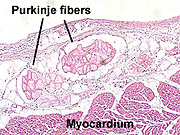
- Identify Purkinje fibers (Fig.
11-4) of the heart's conduction system near the endocardium and epicardium.
What is unusual about the
embryonic origin of Purkinje fibers in light of how they function?
The heart valves
are next. |