Heart valves must be shaped
so as to exactly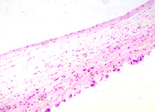 fit together and prevent backflow of blood. If congenitally abnormal
or misshapen due to infection or other problem, regurgitation of
blood back through the valve will occur and this is detectable as a
murmur.
fit together and prevent backflow of blood. If congenitally abnormal
or misshapen due to infection or other problem, regurgitation of
blood back through the valve will occur and this is detectable as a
murmur.Examine the histology
of heart valves in micrographs (Fig. 11-6).
Study of the circulatory system
outside the heart includes the arteries and veins, their smaller
branches, and the microvasculature between the arterial supply and
the venous system.
 The
relative thicknesses and the general shapes of these vessels in
transverse section is shown in the diagram. The
relative thicknesses and the general shapes of these vessels in
transverse section is shown in the diagram.
Arteries and Arterioles
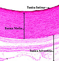
Examine a section of the aorta (slide
100,
115 and
146) and identify (click image to expand)
- The three basic layers or tunics (intima,
media, and adventitia) of this large vessel
- Note their distinctive features
(Figs. 11-7 through 11-10).
What are the locations of the
vasa vasorum, elastin sheets, smooth muscle fibers, the endothelium?
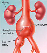
Clinical note: Degeneration of
components such as elastic fibers in the tunica media of arteries
weakens the arterial wall and can lead to blood leakage into or
through the wall (a “dissected” artery) or to formation of a
circumscribed bulge called an aneurysm. Either type of defect can
have disastrous results in a major artery such as the aorta.
Now for muscular
and elastic arteries. |