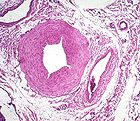
On the slide of heart (slide 65)
examine the coronary artery as an example of a large muscular
artery.
- The tunica intima is relatively
thick (which may indicate an early stage of atherosclerosis) and
- The internal elastic lamina is
prominent.
- Note that tunica media smooth
muscle is more abundant than in the aorta.
- Note also how the tunica
adventitia merges into surrounding CT of the heart.
Why are internal elastic lamina
and the intima commonly folded like an accordion in muscular
arteries?
 Small
muscular arteries (Fig. 11-11) can be seen on many slides in your
collection. Examine muscular arteries of different sizes in areolar
CT (slide 92). Small
muscular arteries (Fig. 11-11) can be seen on many slides in your
collection. Examine muscular arteries of different sizes in areolar
CT (slide 92).
- Note the frequent location along a
vein and small nerve.
- Identify the three layers in the
artery walls.
How does the distribution of
elastin differ between elastic and muscular arteries?
Arterioles and
capillaries. |