 The
basic and most common stain used in histology is actually a
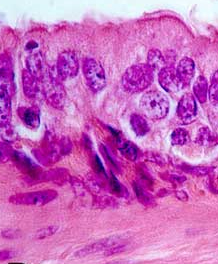
combination of two stains, hematoxylin and eosin (H&E). The
basic and most common stain used in histology is actually a
combination of two stains, hematoxylin and eosin (H&E).
The charge of the tissue constituents,
proteins, nucleic acids, lipids, etc, determine which stain will
preferentially bind.
- Basic stain (e.g. hematoxylin)
contains positive charges (cationic) and binds to negatively
charged (basophilic) substances, such as nucleic acids.
- Acid stain (e.g. eosin)
contains negative charges (anionic) and binds to positively
charged (acidophilic) substances, such as proteins.
Most of the slides in your
collection, and most of those shown in Junqueira, are stained with H&E. This
is by far the most routine stain used in hospital pathology labs
and will be the same stain used for the tissues you will see
next year in pathology.
Other commonly used stains bind
preferentially to specific cellular or tissue
constituents. |