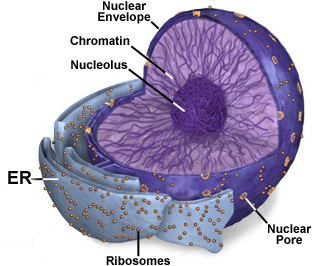
| The nucleus, generally the largest
and most easily seen organelle in a cell, is filled with basophilic
chromatin. Chromatin consists of DNA associated with various
proteins and RNA. In routinely stained slides, nuclei can show
regions of euchromatin and heterochromatin in areas of high and low
gene activity respectively.
 In
cells synthesizing protein very actively, the specific sites in
chromatin with the genes for rRNA become more intensely stained
because of the rRNA precursors accumulating there and are seen as
nucleoli. Examine TEM picture of nuclei (Fig. 3-2).
Examine the nuclei of neurons and other cells on
slide 71, of liver
cells (slide 141), and cells of the pancreas (slide 44), locating
the areas mentioned above. In
cells synthesizing protein very actively, the specific sites in
chromatin with the genes for rRNA become more intensely stained
because of the rRNA precursors accumulating there and are seen as
nucleoli. Examine TEM picture of nuclei (Fig. 3-2).
Examine the nuclei of neurons and other cells on
slide 71, of liver
cells (slide 141), and cells of the pancreas (slide 44), locating
the areas mentioned above.
Choose any three cell nuclei on the pancreas slide and draw
them here, indicating especially the areas of euchromatin and
heterochromatin.
Compare and contrast the staining properties and activities of
“heterochromatin” and the nucleolus.
The nuclear envelope consists of two membranes:
- The outer is continuous with the
ER and the inner is associated with a layer of proteins (the
nuclear lamina)and heterochromatin.
- Passage of ribonucleoprotein
particles through the envelope into and out of the nucleus is
regulated by nuclear pores. Examine these in the TEM of
Fig 3-5
and in the "freeze-etch" EM preparation of Fig. 3-6.
How does DNA folding in
chromatin involve "nucleosomes" and how does DNA folding differ
between euchromatin and heterochromatin?
List 3 macromolecules that
leave the nucleus through nuclear pores and 3 that enter through the
pores.
Cell division. |