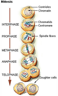
 Examine
the onion root tip preparation (slide 93) which shows
mitosis in
nicely aligned cells better than mitotic cells can be seen in any
animal tissue. Find examples of all stages of mitosis: prophase,
metaphase, anaphase, telophase (Fig. 3-14). Examine
the onion root tip preparation (slide 93) which shows
mitosis in
nicely aligned cells better than mitotic cells can be seen in any
animal tissue. Find examples of all stages of mitosis: prophase,
metaphase, anaphase, telophase (Fig. 3-14).
In sectioned animal tissues, specific
stages of mitotic cells are very difficult to identify. Mitotic
cells have very condensed chromatin and can be located easily only
in certain tissues of adult organs, including among the epithelial
cells of endometrial glands in the growth phase (slide
80 and
83) of certain lymphoid tissues (slide 136),
and in the crypts of the small intestine (slide
37). Fig. 3-17 shows examples of these.
|
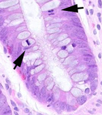 |
To the left is a section of
normal, healthy colonic mucosa.
- The arrows indicate
two cells in different stages of mitosis.
- Can you tell what
phase each is in?
|
Why are we looking at a slide
of onion cells in a human histology lab?
Why are the stages of mitosis
hard to make out in dividing cells present in routinely prepared
sections of adult tissues?
Now for cellular secretion, membranes and
organelles. |