Anatomy A215 Virtual
Microscopy
|
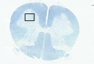 |
In the case of the spinal cord, the gray and white matter are
readily distinguishable. The gray matter has the appearance of a fat
letter 'H' when the spinal cord is viewed in cross section. The
white matter surrounds the gray matter in the case of the spinal
cord. Large motor neurons are characteristic of the anterior horns
of the spinal cord. |
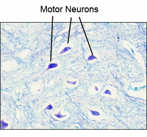 |
In this SPINAL CORD slide,
locate a large motor neuron in the anterior horn, and
identify the following features of it:
- Cell body (or perikaryon or soma)
- Nucleus with a distinct nucleolus
- Chromatophilic
substance (or Nissl bodies), dark granular material within the
cytoplasm
- Nerve cell processes of two types:
- Dendrites and
- An axon (arising from a lightly staining, cone-shaped
area known as the axon hillock)
- Click here for a detailed image of the spinal cord.
- Click here for a detailed image of a motor neuron.
|
A215 Home Page
| Virtual Microscopy
Table of Contents
|