Anatomy A215 Virtual
Microscopy
|
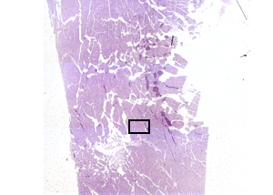 |
This virtual microscope HEART slide is of a piece of human heart. It
includes not only the muscle of the heart, but also the outer
covering known as the epicardium. The little box in the image to
the left indicates where the photograph below came from.
This area is of cardiac muscle, just beneath the epicardium. |
|
 |
The wall of the heart has three
layers which can be distinguished histologically:
- The thin, outer epicardium.
- The thick myocardium, containing an interlacing network of
cardiac muscle fibers. Note in particular the
intercalated discs, which are connections between cardiac
muscle cells.
- The thin endocardium, the innermost layer of the heart.
|
A215 Home Page
| Virtual Microscopy
Table of Contents
|