Anatomy A215 Virtual Microscopy
Blood Vessels


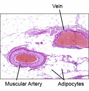
Blood vessels are of three main types: arteries, capillaries, and veins. Arteries and veins have a 3-layered pattern to their walls. 
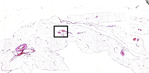
The little box in the image to the left indicates where the picture below came from. There are plenty of little blood vessels in this slide so don't feel you have to stick just with ones we chose for demonstration.
 

Arteries carry blood at high pressures, and therefore must have thick walls. In all arteries, the tunica media is the thickest layer, but its composition varies among the three general classes of artery.

Several blood vessels are scattered throughout the mesentric fat of this MESENTERY slide. Use this slide to identify the different layers, or tunics, of a blood vessel’s wall (the Latin noun"tunica" (covering or coat) is used for this type of layer):
  • The tunica externa (sometimes called tunica adventitia) is the outermost layer.
  • The tunica media contains circularly arranged smooth muscle cells and elastic fibers. The ratio of these components varies among different types of vessels.
  • The tunica intima, the innermost layer, consists of connective tissue and a surface of simple
    squamous epithelium forming the lining of the vessel.
  • Click here for a detailed view of a muscular artery.
  • Click here for a detailed view of an arteriole and venule.
See this slide with the virtual microscope

I don't see the virtual slide, I must need the download.


   A215 Home Page | Virtual Microscopy Table of Contents