
General
and Systemic Histopathology, C601&C602
Slide 77: Lymph node with tuberculosis
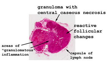 |
Although this slide is
a bit faded, you should be able find the areas of granulomatous inflammation.
In some areas there are well developed granulomas with giant cells.
See this slide with the
virtual microscope. |
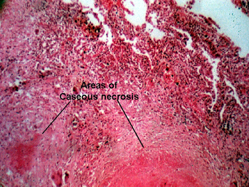 |
This is a pulmonary hilar
lymph node showing a granuloma and many giant cells. It is from the person
that also gave you slide #76, and demonstrates the body's reaction to TB.
As with slide #76, the overall pattern of the granuloma is the same. |
Back
to Home
|