General
and Systemic Histopathology, C601&C602
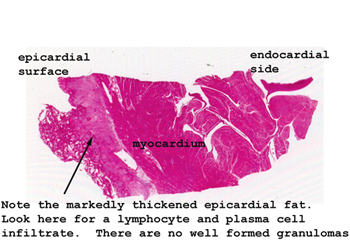
Slide 78: Tuberculous pericarditis

|
Here you will see an
unbelievable thickened pericardium with a marked chronic inflammatory infiltrate.
The exudate is partially "organized," but no well defined granulomas are present.
We know this was tuberculosis because of the history and positive autopsy
cultures.
See this slide with the
virtual microscope. |

|
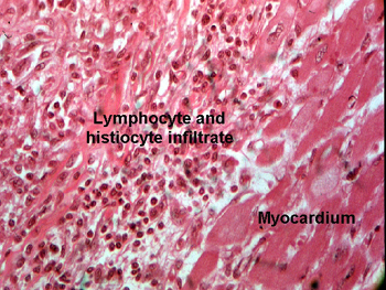
This slide does not
show well developed granulomas, rather a marked chronic inflammatory infiltrate
with a large amount of granulation tissue. By the way, be sure you know the
difference between "granuloma" and "granulation tissue," even if the two
seem to blend together here. In this slide there are many plasma cells along
with the angioblasts and fibroblasts in, and on, the surface of the epicardium.
Histologically it's not really possible to make a diagnosis of TB from what
you have. We know it because of the patient's history and a successful culture.
I am not trying to fool you or give you something you can't diagnose, rather
I am showing you how it can look. |
Back to Home
|