
General
and Systemic Histopathology, C601&C602
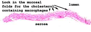
Slide 137: Gallbladder with cholesterolosis
|
Look in the little
mucosal folds for the lipid laden histiocytes.
See this slide with the
virtual microscope. |
|
It helps to have an
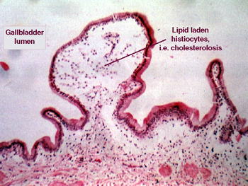
idea of normal gallbladder morphology to see what is wrong with this picture.
You will see somewhat enlarged mucosal folds of the gallbladder, and in many
there will be an infiltrate of foamy histiocytes. There is very little inflammation
of the acute or chronic type here, and if there is any at all, it will be
found in the muscular wall and serosal fat. This is a very common and benign
process, and very likely is the starting point for some types of gall stones. |
Back to Home
|