
General
and Systemic Histopathology, C601&C602
Slide 152: Ruptured Epidermal Inclusion
Cyst of Skin
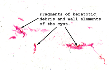 |
OK, there isn't much
to this one, but it's pretty common for us in the clinical lab to receive
a specimen that looks like this. Generally the surgeon just pulls
out fragments of the wall of the cyst and bits of its contents. Take
note of the lining and keratotic matter.
See this slide with the
virtual microscope. |
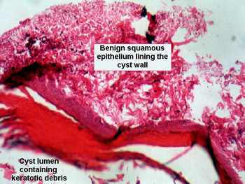 |
These are very common
lesions of the skin, and may result from the implantation of a small fragment
of viable epidermis into the deeper layers of the dermis or subcutaneous
tissues. The lesion is just as it sounds, a cyst composed of epidermal
elements. The cyst lining is composed of benign squamous epithelium, and
the lumen eventually fills with shed keratotic matter. If these rupture,
the keratin debris is considered "non-self" by the immune system, and there
follows a significant and often rather painful foreign body type inflammatory
reaction. Because of the swelling associated with the reaction to the keratin
from a ruptured epidermal inclusion cyst, the person considers the lesion
to have "grown" dramatically almost overnight. As you can guess, this is
very worrisome, and most folks come right in to have it looked at, thinking
it must be cancer because of the rapid "growth." |
Back
to Home
|