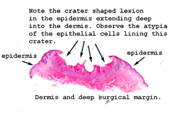
This is an absolutely
classic appearance for this lesion; at least in two dimensions. Deep
central crater, lined by highly atypical cells. As always, start
at the very edge of the biopsy to get oriented as to more or less normal
skin. Take note of the degree of actinic degeneration within the
dermis. Then move to the lesion itself. Stay on low power to
get the overall feel of this condition.
This is an important
lesion to know, it's very common and almost always on sun exposed parts
of the body. The backs of the hands and helix of the ears are very common
sites. This lesion is difficult to distinguish from its more aggressive,
invasive squamous cell carcinoma cousins. Although long thought to be some
benign variant or transitional form of squamous tumor, we now consider
this to be a low grade, locally invasive form of squamous cell carcinoma.
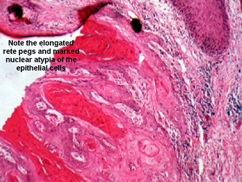
Note the "cupped shaped" nature of the lesion. To see this important diagnostic
feature, you will need to look at the slide on a white background. Other
important microscopic features include keratin "pearls," and an advancing
margin along the base of the lesion. There is no question, the cells of
this tumor look a good bit wilder than they behave, still removing is the
way to go.