
General
and Systemic Histopathology, C601&C602
Slide 139: Acute Appendicitis
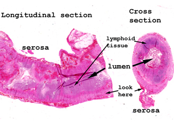 |
Here you have two sections
of the appendix. The one on the left is a longitudinal section in
which the lumen is not well defined. It contains lots of necrotic
debris. The section on the right is a little easier to understand
as a hollow organ. Still the lumen is partially obliterated by necrotic
debris and inflammatory material.
Start reviewing this slide
in the lumen and work your way methodically to the serosal surface.
Pay attention to all the elements and make notes on what you see.
See this slide with the
virtual microscope. |
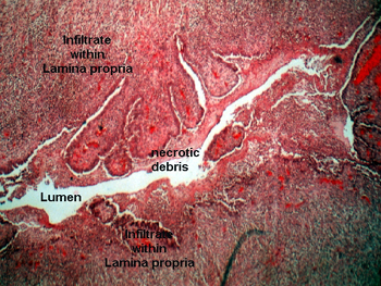 |
In this slide, the mucosa
of the appendix is largely missing and there is a profound acute inflammatory
infiltrate in the lamina propria. There is also a lot of necrotic debris
in the lumen of the organ. This is a difficult slide because the acute
inflammatory infiltrate is intermixed with the normally occurring lymphoid
tissue of the appendix. Remember that in the healthy state you would find
many lymphoid aggregates in the lamina propria of the appendix, and throughout
the length of the bowel for that matter. You may also see some newly forming
granulation tissue on the serosal surface. As far as that goes, your best
shot at seeing the constituents of the acute infiltrate will be in the
serosal surface itself. In some of the slides there is marked lymphoid
hyperplasia in the lamina propria (a finding you might expect), so it is
probably best to steer away from lumen and the centrally located portions
of this tissue for right now. |
Back
to Home
|