
General
and Systemic Histopathology, C601&C602
Slide 97: Adrenal tuberculosis
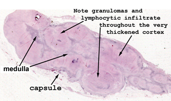 |
Just by looking at the
tissue on the slide, you can see the marked alteration in the normal appearance
of this adrenal gland. When disseminated, TB often goes to the adrenals.
Even without the microscope you can see the extensive areas of caseous
necrosis throughout the cortex.
See this slide with the
virtual microscope. |
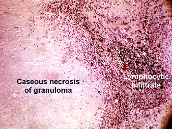 |
This is a pretty washed
out slide, do your best, but don't waste a lot of time. As you will see,
there is practically no adrenal gland left. We would be hard put to even
tell the organ had it not been for its location at the time of autopsy.
Observe the granuloma with caseous necrosis at its center. There are few
giant cells, but the principal remaining inflammatory pattern is of non-specific
chronic inflammation. You will see lymphocytes and plasma cells comprising
the majority of the inflammatory pattern. |
Back
to Home
|