
General
and Systemic Histopathology, C601&C602
Slide 172: Lung with Interstitial pneumonia
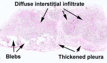 |
This slide a lot going on. There is
evidence of previous inflammation of the pleura, as noted by the scarring,
plus changes of emphysema and then the interstitial pneumonia.
See this slide with the
virtual microscope. |
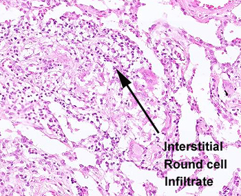 |
Compare this slide with the case
of lobar pneumonia, slide 120. Here you will see the infiltrate is
interstitial (not within the alveolar air spaces), and consists almost
exclusively of lymphocytes. What infectious agent is likely to result in the
picture seen in this slide?
|
Back to Home
|