
General
and Systemic Histopathology, C601&C602
Slide 135: Lung with features of DIC
|
The best bet here is
to look in the capillary sized vessles for the diagnostic changes. You're
looking for thrombi composedonly of protein. If you see what looks
like thrombi with lots of RBC's in them, it's not what we're looking for.
See this slide with
virtual microscope. |

|
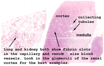
There are two pieces
of tissue on this slide, and obviously this is the lung. The pathological
features are the same for both the kidney and lung. With disseminated intravascular
coagulation (DIC), the person experiences "run away" intravascular coagulation.
As you might expect, there will be small thrombi in vessels throughout the
body. This becomes an ischemic disease on the cellular level. People bleed
with this condition because of the breakdown of the small vessels and the
consumption of the clotting agents. Causes are gram negative sepsis, massive
trauma and obstetrical disasters, just to name a few. This condition never
just arises out of the blue, it is always a complication of something else. |
Back to Home
|