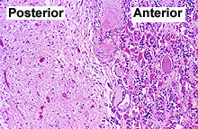
 Examine
the posterior pituitary on slide 122 and identify parts of the
microvasculature, non-myelinated axons, and the support cells called
pituicytes (Fig. 20-11). The non-myelinated axons present are
difficult to see without special stains. Examine
the posterior pituitary on slide 122 and identify parts of the
microvasculature, non-myelinated axons, and the support cells called
pituicytes (Fig. 20-11). The non-myelinated axons present are
difficult to see without special stains.
Where are the two hormones synthesized
which these axons secrete and how is the secretion controlled?
To what CNS cells are
pituicytes of the neurohypophysis analogous?
Clinical note: Diabetes
insipidus is a disorder in which the neurohypophysis fails to
secrete vasopressin/antidiuretic hormone in response to normal
stimuli. The disorder involves excessive thirst, excessive water
intake, and excessive urination and can be treated by injection of
vasopressin or similar drugs.
 Pineal
gland -- small neurosecretory organ in brain with axons
communicating to other parts of the brain. The pineal is a
photoreceptor organ in primitive vertebrates. Pineal
gland -- small neurosecretory organ in brain with axons
communicating to other parts of the brain. The pineal is a
photoreceptor organ in primitive vertebrates.
Examine pineal tissue on slide
106 or
107. Pineal tissue can be identified from surrounding brain tissue
by the presence of pineal sand (Fig. 20-24), concretions of
mineralized organic matrix of unknown significance. Note the
abundant vasculature. Identify the small clumps of pinealocytes,
with scant poorly stained cytoplasm and ovoid nuclei containing
nucleoli. The fibrous material surrounding all the cells is
primarily neuropil, not collagen.
What function is primarily
associated with the pineal gland?
“Pineal sand’ has medical relevance
because radiologists find that it is a good marker of the brain’s
midline.
Next is the
thyroid. |