 Examine
the trichrome-stained section of prostate gland on
slide 95.
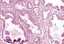
Identify the tall epithelial cells with lightly stained "foamy"
cytoplasm at the apical ends (Figs. 21-13 and 21-14) and in the stroma notice
the smooth muscle fibers (red) mixed with dense connective tissue
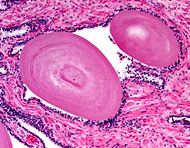
(blue). In the lumen identify the calcified, proteinaceous
concretions called corpora amylacea (Fig. 21-16). Examine
the trichrome-stained section of prostate gland on
slide 95.
Identify the tall epithelial cells with lightly stained "foamy"
cytoplasm at the apical ends (Figs. 21-13 and 21-14) and in the stroma notice
the smooth muscle fibers (red) mixed with dense connective tissue
(blue). In the lumen identify the calcified, proteinaceous
concretions called corpora amylacea (Fig. 21-16).
What explains the poor staining
properties of the cells of the prostate?
What may cause corpora amylacea
to form?
The association between prostate and
urethra can be seen in the specimen on
slide 47, which shows more of
the gland surrounding urethra, although the latter has been split
and appears on the "edge" of the section, unlike its disposition in
Fig. 21-15. Notice the difference between the glandular tissue near
the urethra and the glands throughout most of the organ further from
the urethra.
Clinical note: Prostate glands
provide urologists with plenty of work by being prone to three very
common problems: (1) They are the site of chronic, low-grade
bacterial infections. (2) In older men the secretory epithelium very
frequently undergoes benign hyperplasia, resulting in so much tissue
overgrowth that the urethra is often constricted and there are
problems with urination. (3) Adenocarcinoma of the prostate
epithelium is now the most common form of cancer in men.
Speculate why prostate
infections are much more common than infections of seminal vesicles.
Could the same reasons explain
why prostate cancer is so common and seminal vesicle cancer so rare?
The penis and
urethra. |