Ganglia are collections of
cell bodies outside the CNS, for sensory or autonomic nerves.
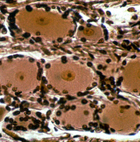
- Outside the spinal cord on slide
9 and 6T, locate and examine a spinal or dorsal root ganglion
(Fig. 9-29a,b). (Another example of a larger dorsal root ganglion
is sectioned on slide 137.)
- Identify the large neurons
(which are sensory),
- The glial satellite cells
surrounding each neuron (Fig. 9-29c,d, and
- The CT covering the entire
ganglion.
What is the difference between
the satellite cells of ganglia and the satellite cells you learned
about in muscle?
Clinical note: The
herpes zoster virus, acquired during childhood chicken pox, can
remain dormant in neurons of sensory ganglia and can be reactivated
in older individuals, migrating along axons to the skin and
producing blisters and a painful rash known as shingles. Although
skin symptoms usually heal within weeks, the pain (post-herpetic
neuralgia) may persist for many months.
Examine an sympathetic ganglion (Fig.
9-29c) on slide 135 and identify the same structures just
found in the sensory ganglion.
- Smaller parasympathetic ganglia will
be found in the wall of the small intestine (slide 37),
esophagus (slide 43), and in some areas of loose CT (slide 45).
What are similarities and
differences between an autonomic ganglion and a spinal ganglion?
Now let's look at
peripheral nerves. |