
General
and Systemic Histopathology, C601&C602
Slide 42: Liver with cirrhosis
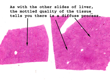 |
Here just looking at
the tissue on the slide, you can see the evolving nodular pattern so characteristic
of cirrhosis.
See this slide with the
virtual microscope.
|
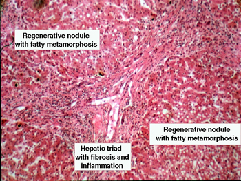 |
This slide shows all
of the features of cirrhosis. You can probably see the degree of nodularity
best by first looking at this slide on a white background before going
to the microscope. Observe the "bridging fibrosis" between the triads and
the loss of definition of the limiting plate of the triads. There is still
much inflammation. Note the "regenerating nodules." These nodules are isolated
from the biliary system and represent an effort by liver to repair damage,
but it's clearly uncoordinated and ineffective. The deranged vascular
flow will ultimately become a major problem for those with cirrhosis. |
Back
to Home
|