
General
and Systemic Histopathology, C601&C602
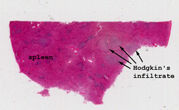
Slide 57: Hodgkin's Disease in Spleen
|
The areas of splenic
involvement are pretty obvious here, but this isn't always the case.
Look at the uninvolved spleen first to get oriented and then focus on the
areas of tumor.
See this slide with the
virtual microscope. |
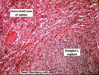
|
The involvement with
Hodgkin's is easy to see in this slide, although this is not always the case.
You should be able to find Reed-Sternberg cells within this splenic implant.
Remember that when trying to identify them, it is essential that they are
in "their proper background." That is to say that they are seen with the
Hodgkin infiltrate consisting of lymphocytes, plasma cells and especially
eosinophils. There are many cells seen in a reactive lymph node that will
mimic the Reed-Sternberg cell. Before identifying anything as one of these
diagnostic cells, be sure it is in its proper setting! |
Back to Home
|