|
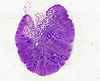
General and Systemic Histopathology, C601&C602 Disorders of Lymph Nodes and Reticuloendothelial System In this unit we are going to look at several aspects of lymph node pathology, both benign and malignant. Remember that the lymph nodes are potentially catching a lot of "floating" and foreign material, so they frequently become secondarily involved in many conditions. Because of this property we are going to look at them in situations you might not at first suspect. Furthermore, we are going to look at situations in which proliferative conditions of the lymphatic system involve organs as an "innocent bystander." There are a number of organs that may not leap to mind as harboring a great deal of lymphatic tissue, but there are. Consider for example the bowel. The most common primary malignant tumor of the small bowel happens to be a malignant lymphoma. Not surprising once one considers the large amount of lymphatic tissue in the lamina propria throughout the small bowel. But before going to the slides and descriptions, here are a couple of suggestions and "helpful rules" for interpreting microscopic sections of lymph nodes. By now, it should be
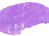
ingrained in your thinking to look at a slide on a white background before
putting it on the stage of the microscope. It's doubly important in the
case of lymph nodes. This visual assessment will often provide much information
about the extent of replacement or involvement of the node with whatever
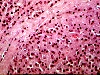
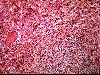
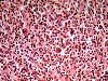
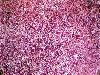
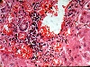
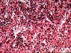
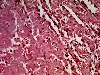
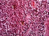
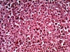
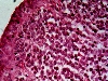
process afflicts it. Also, when assessing a lymph node, keep in mind the node
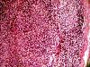
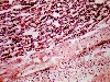
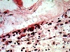
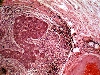
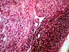
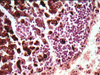
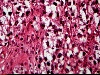
should have a characteristic microscopic architecture. That is, a capsule,
subcapsular sinuses, cortical tissue and cortical sinuses, and so forth. We
always look at all aspects of the lymph node, and assess the degree of effacement
of the nodal architecture. That is to say the degree of replacement of the
expected nodal architecture with whatever disease process is going on. The
description and understanding of what is meant by "the either complete or
partial effacement of a lymph node" is not only crucial to recognizing the
underlying disease process, it is crucial to your grade. Questions about this
term will come up on the test, and you can bet when you are asked to diagnose
a slide with a malignant lymphoma, I'm going to looking for the appropriate
terminology in your write-ups. Although there are a number of slides
in this unit, I'd recommend taking a little extra time, and possibly going
over your observations with a friend. Good luck.
Previous Laboratory | Next Laboratory | Table of Contents
Copyright 1998, the Trustees of Indiana University
|