
General
and Systemic Histopathology, C601&C602
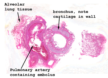
Slide 108: Lung with pulmonary embolus
|
There are several pieces
of tissue on the slide. The one with the pulmonary embolus should be obvious.
The hole in the middle of the clot is an artifact of sectioning. It
was in fact solid but partially chipped out when it was sectioned.
See this slide with the
virtual microscope. |
|
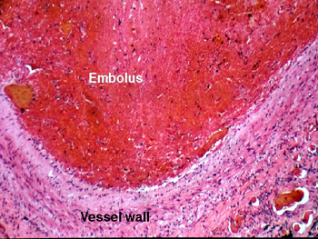
There is nothing real
fancy in this slide. It is a pulmonary embolus. Be sure you know the difference
between a thrombus and an embolus, as well as the sorts of things that can
potentially embolize. For that matter, be sure you know the factors that
contribute to the formation of a thrombus. Remember there are differences
in formation on the venous and arterial sides of the circulation. Here's
an interesting problem I almost always ask about on the written exams:
paradoxical embolization
. Know what it is? |
Back to Home
|