Anatomy A215 Virtual
Microscopy
| Chapter 2: Epithelial and
Connective Tissues |
|
|
| Epithelia form layers covering
surfaces or lining cavities; they often fold inward to form glands.
They may consist of a single layer of cells (simple) or more than
one layer (stratified); they lie on a very thin basement membrane.
The side of an epithelial layer opposite the basement membrane is
called its free surface.
Connective tissues consist of three basic components: cells, fibers,
and ground substance. The proportions and nature of these three
components vary in the different connective tissues depending on
their location and function. |
|
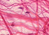 |
In this slide of
AREOLAR CONNECTIVE TISSUE (often called simply “loose connective”
tissue), observe the loose and unorganized arrangement of the
collagen fibers.
Click for slide page. |
|
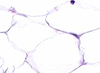 |
Adipose tissue is a form of connective tissue consisting largely of
fat cells (adipocytes). View this slide of MESENTERY to see an
example of adipose tissue. Click for slide page. |
|
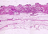 |
In this section of SKIN, you can see dense irregular connective
tissue. Examine the tissue deep to the darkly stained epithelium; note how the components are arranged randomly.
Click for slide page. |
|
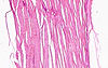 |
In this slide of TENDON, a good example of dense regular
connective tissue, note that the fibers have an orderly and linear
arrangement.
Click for slide page.
|
A215
Home Page
|
Virtual Microscopy
Table of Contents
|